Spotters A3
Slide 2
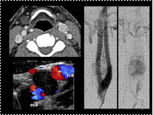
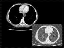
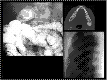
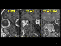
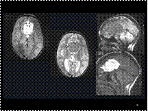
Carotid Body Tumor
Slide 3
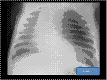
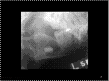
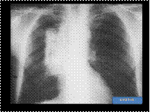
CLE
Slide 4
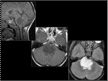
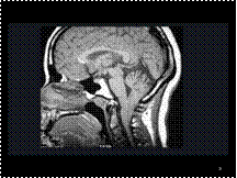
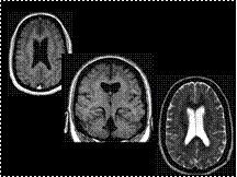
Brainstem Astrocytoma
Slide 5
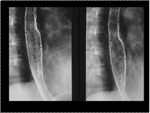
Esophageal Duplication Cyst
Slide 6
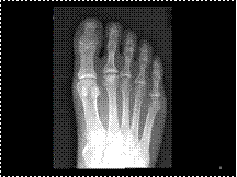
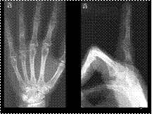
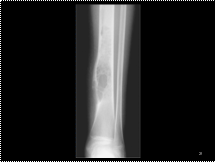
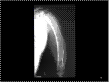
Freiberg oteochondritis
Slide 7
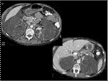
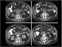
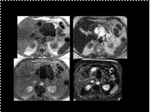
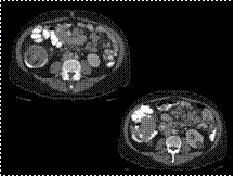
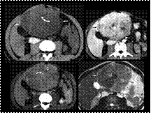
CT of pancreas divisum….Incidental note is made of autosomal dominant polycystic kidney disease in this patient
Slide 8
Sprengel's Deformity
Slide 9
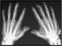
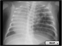
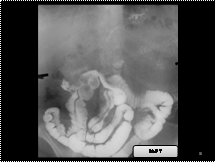
Gardner syndrome ---Upper GI examination shows a polypoid filling defect in the proximal...... duodenum. Radiograph of the right chest shows multiple sessile sclerotic osteomas. CT of the mandible shows multiple benign osteomas (arrows).
Slide 10
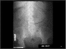
Retroperitoneal fibrosis
Slide 11
Right AA
Slide 12
Gallstone ileus
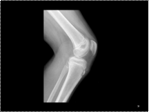
Slide 13
Osgood Schlatter
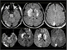
Slide 14
PRES
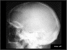
Slide 15
pagets
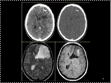
Slide 16
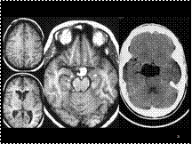
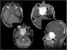
diagnosis is oligodendroglioma….
differential diagnosis... includes a malignant astrocytoma or a glioblastoma
Slide 17
Inverted papilloma
Slide 18
Candida Esophagitis
Slide 19
Chiari I Malformation
Slide 20
PSORIATIC ARTHROPATHY
Slide 21
Lipoma with Callosal Agenesis
Slide 22
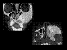
Odontogenic keratocyst in a 13-year-old boy. An abnormality was seen incidentally on conventional radiographs obtained for planning of orthodontic treatment. (a) Lateral oblique radiograph shows an ellipsoid, expansile, multilocular, corticated, lucent lesion occupying the anterior two-thirds of the left ramus with an impacted third molar crown displaced inferiorly within the lesion. The mandibular canal appears to be displaced inferiorly as well. (b) Posteroanterior radiograph shows buccal (lateral) displacement of the third molar by the lesion. The patient underwent en bloc resection. The differential diagnosis includes dentigerous cyst, odontogenic keratocyst, and ameloblastoma.
Slide 23
Enchondroma
Multiple enchondromas = Ollier's Dz
Multiple enchondromas + hemangiomas of soft tissue = Maffucci syndrome
Slide 24
Ruptured Dermoid
Slide 25
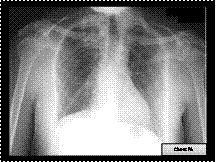
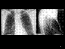
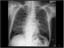
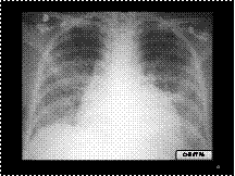
lymphangitic carcinomatosis
Slide 26
Band Heterotopia
Slide 27
Admantinoma
Slide 28
FD
Slide 29
Post RT
Slide 30
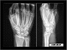
Lunate Dislocation
Slide 31
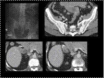
Serous cystadenoma pancreas
Slide 32
Emphysematous Gastritis
Slide 33
Fat containg lobulated mass with in the colonic lumen : Intussusception
Slide 34
CCAM
Slide 35
FLC
Slide 36
ASCARIASIS

Slide 37
Cerebral autosomal dominant arteriopathy with subcortical infarcts and leukoencephalopathy (CADASIL)
Slide 38
FD
Slide 39
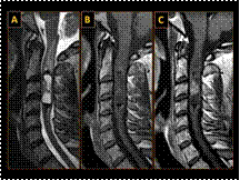
Cellular Ependymoma—Cervical Cord
Slide 40
PCP
Slide 41
Chordoma