Spotters B4

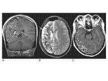
Slide 2
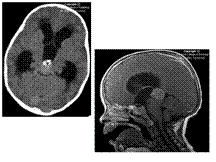
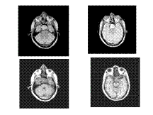
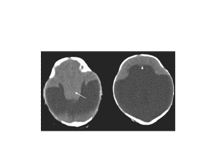
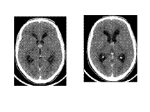
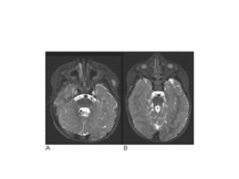
Pineal germinoma
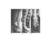
Slide 3
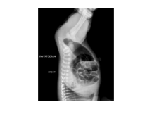
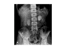
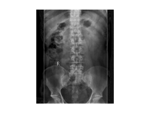
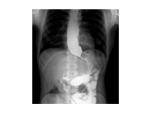
Organoaxial volvulus
Slide 4
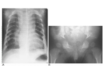
PAH
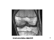
Slide 5
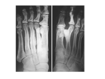
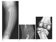
Osteiod osteoma
Slide 6
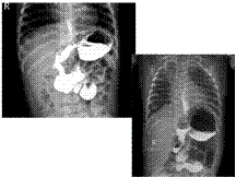
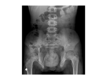
Imperforate anus
Slide 7
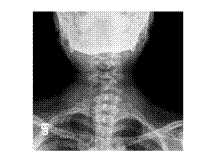
Dextrocardia with situs inversus
Slide 8
Pituitary macroadenoma
Slide 9
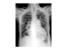
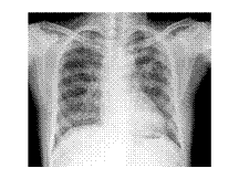
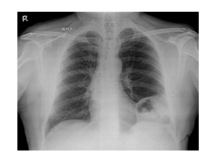
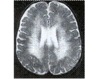
miliary
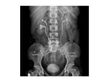
Slide 10
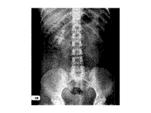
Bladder extrhopy
Slide 11
Alobar holoprosencephaly Figure 70.10 CT brain of this infant shows that the cerebral hemispheres have failed to form and there is no interhemispheric fissure or corpus callosum. Instead there is a thin pancake of cerebral tissue crossing the midline anteriorly (arrowhead) and a single holoventricle continuous with a large dorsal cyst. The midbrain and deep grey structures are fused into a single indiscriminate mass (arrow).
Slide 12
Melorheostosis (two patients). There is dense irregular cortical bone: this is sometimes described as the ‘flowing candle wax’ appearance. One patient demonstrates a ‘ray’ distribution (asymmetrical changes).
Slide 13
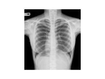
Asphyxiating thoracic dystrophy. (A) Narrow thorax and short ribs. (B) Horizontal acetabular roofs and pronounced medial spurs, less pronounced laterally (‘trident’ appearance).
Slide 14
Sturge–Weber syndrome. (A) Coronal T1 post-contrast image shows an enhancing pial angioma overlying the right cerebral hemisphere which is atrophic. The right choroid plexus is enlarged. Foci of signal hypointensity within the gyri and adjacent white matter are due to calcification. (B) Axial T2-weighted image shows in addition prominent superficial cortical veins and ependymal veins (arrows). (C) Axial post-contrast T1-weighted image shows bilateral choroidal angiomas (arrows) in addition to the pial angioma
Slide 15
Duplex system
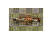
Slide 16
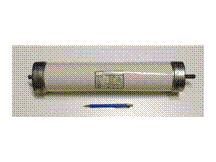
Stationary anode xray tube
Slide 17
Vas deferans calcification
Slide 18
Colloid cyst
Slide 19
b/l pujo
Slide 20
DH
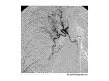
Slide 21
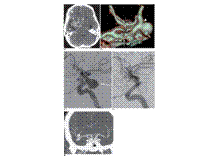
Aneurysms on CTA. (A) CT shows acute subarachnoid blood in interhemispheric and right Sylvian fissures (short arrows) and a clot in the right side of the chiasmatic cistern extending into the temporal lobe (open arrow). Within this is a rounded area of lower density, suggesting an aneurysm (long arrow). There is also an acute right subdural haematoma (arrowheads). (B) CTA viewed from above, behind and the left shows a large right distal ICA aneurysm arising at the level of the posterior communicating artery (not visible as anatomically hypoplastic). The neck is clearly shown (arrowhead) and there is a small lobule (short arrow) on the fundus possibly indicating the site of rupture. Note the left posterior communicating (long arrow) and anterior communicating arteries (open arrow). The decision to treat by endovascular coiling was based on this examination. (C, D) Lateral right ICA arteriograms from coiling procedure, immediately before and after occlusion of the aneurysm. Note confirmation of aneurysm anatomy as shown on CTA, including terminal lobule (arrow). (E) Coronal MIP from CTA in a different patient with a complex right middle cerebral artery aneurysm. Note the anatomical detail of separate lobules and artery arising from the neck of the superior aneurysm (arrow)
Slide 22
Lipomyelomeningodysplasia. Sagittal (A) T1- and fast spin-echo (B) T2W and coronal (C) T2W images showing the lipoma, low position of the spinal cord and a cavity in the distal spinal cord
Slide 23
Segond fracture of the knee. Coronal proton density-weighted MRI demonstrates avulsion of the bony insertion of the iliotibial band (arrow). Avulsions of the lateral collateral ligament complex have a close association with ACL injury
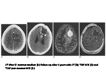
Slide 24
Oligodendroglioma. CT after IV contrast medium (A) shows a large left frontal tumour that involves the cortex. It is predominantly solid with irregular enhancement, but there are also cysts and coarse calcification. Follow-up after 2 years with CT (B), T2W MRI (C) and T1W post-contrast MRI (D) shows more extensive cyst formation and calcification than on the first scan. The calcification is much less apparent on MRI and appears as nonspecific low signal areas. Posterior infiltration of the tumour is, however, best seen on MRI (C). Note that the patient had undergone a left frontal craniotomy after the first CT
Slide 25
Bronchial a embolization
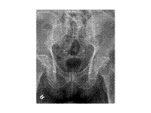
Slide 26
b/l sacroilitis
Slide 27
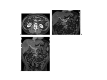
Metastatic carcinoid tumor. A: Transaxial computed tomography shows a lobulated soft tissue mass (arrow) with punctate central calcifications at the root of the small bowel mesentery. Strands of soft tissue density radiating from the mass toward the small bowel loops are indicative of desmoplastic response to the tumor. B: Coronal image also demonstrates the characteristic features of the mass. C: Coronal maximum intensity projection (MIP) image shows engorgement of the mesenteric veins due to partial obstruction by the mass. Note the large hepatic metastasis (arrowhead)
Slide 28
Ca esophagus
Slide 29
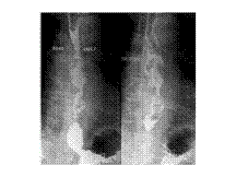
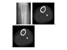
Enchondroma: CT appearance. A: AP radiograph of the left proximal calf shows a proximal diaphyseal lesion with intralesional calcifications and slight expansile remodeling. B: and C: Axial CT images show the central ring and arc like calcifications and peripheral lucency, as well as the mild circumferential endosteal scalloping
Slide 30
Achalsia cardia
Slide 31
STAPHYLOMA -ENLARGEMENT OF RT GLOBE WITH OUTWARD BULGING OF SCLERA ON THE TEMPORAL SIDE
Slide 32
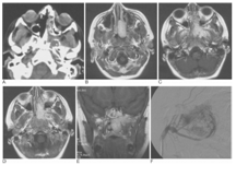
Juvenile angiofibroma in a teenager with epistaxis. (A) This axial CT was the first study performed in this patient. The critical finding is widening of the pterygopalatine fissure on the left side. This is virtually diagnostic of an angiofibroma. (B–E) Contrast-enhanced (gadolinium) T1-weighted MRIs. (B) Axial image revealing an enhancing, well-defined mass in the nose. Clinically this was visible from an anterior view through the nose and a posterior view with a mirror in the nasopharynx. (C) Axial image. Note the lateral extension of the tumour towards the infratemporal fossa. (D) Axial image. More superiorly the tumour is less well defined and more infiltrative. Note the extension into the pterygopalatine fissure is visible but more easily seen on CT. (E) Coronal image revealing the mass at the level of the posterior choana with its lateral extension. This is an essential observation for surgical planning. (F) Angiography. Lateral superselective injection into the maxillary artery shows the highly vascular nature of the tumour. This investigation was performed before therapeutic embolization and subsequent surgical resection
Slide 33
Ch pancreatitis
Slide 34
Thyroid mass
Slide 35
Staghorn calculus
Slide 36
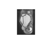
Hydrocolpos. T2-weighted parasagittal MRI showing a markedly distended vagina in a neonate with fluid also seen superiorly in a less distended uterine cavity, seen posterior to the bladder
Slide 37
Blount's disease. (A) Plain radiograph, (B) coronal CT and (C) 3D CT reconstruction (posterior view)
Slide 38
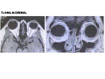
Child with Joubert's syndrome. (A) Typical batwing appearance to the fourth ventricle (arrow) and (B) prominent superior cerebellar peduncles with failure of the normal midline decussation (arrow). This gives the typical ‘molar tooth’ appearance. The midbrain is hypoplastic in this condition
Slide 39
capacitor
Slide 40
PELIZAEUS –MERZBACHER DISEASE
Slide 41
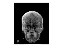
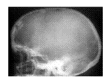
Hyperostosis frontalis interna