Spotters 10

Slide 2
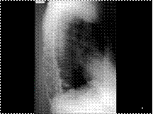
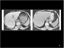
FUNGUS BALL & TB
Slide 3
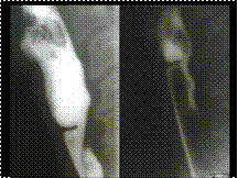
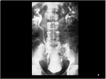
1Retrocaval ureter
Slide 4
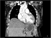
RHD
Slide 5
FD
Slide 6
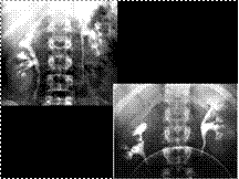
H SHAPE VERT SICKLE CELL ANEMIA
Slide 7
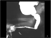
ESOPHGEAL WEB
Slide 8
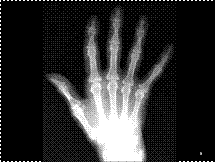
HYPER PTH WITH BROWN TUMOR
Slide 9
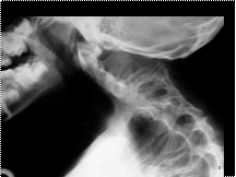
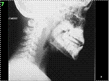
KLEIPPEL FEIL
Slide 10
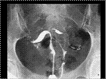
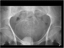
PUV
Slide 11
A DISSECTION STAN A
Slide 12
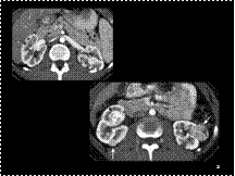
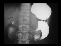
RP ABSCESS
Slide 13
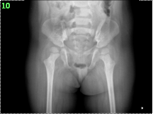
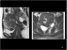
SPINAL DYSRAPHISM R HIP DISLOCATION
Slide 14
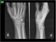
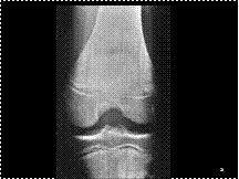
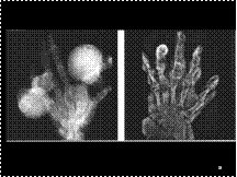
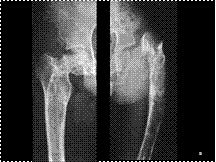
SCAPHOID #
Slide 15
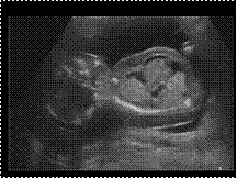
Fetal hydrops. Longitudinal view of a fetus with skin oedema, ascites and a hydrothorax.
Slide 16
Congenital uterine abnormalities HSG images. (A) Bicornis bicollis (single vagina, two cervices, two separate uterine horns).
Slide 17
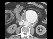
Leiomyosarcoma of the inferior vena cava (IVC).
Slide 18
1Ureteral tuberculosis & Renal tuberculosis. Contrasted CT shows a dilated infundibulum (curved arrow) proximal to a stricture, thickening of the proximal ureter wall (arrow) and renal abscess with peripheral calcifications (arrowheads).
Slide 19
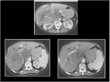
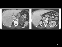
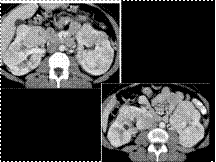
Adrenal tuberculosis. A: Computed tomography reveals a densely calcified right adrenal (curved arrow) and a heterogeneous left adrenal mass (arrows) in this patient with disseminated tuberculosis. B: More inferiorly, both adrenals show heterogeneous, somewhat low-attenuation small masses (arrows). The patient was addisonian.
Slide 20
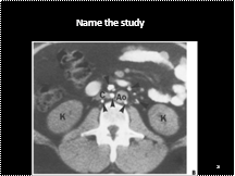
lymphangiography, showing normal distribution of retroperitoneal lymph nodes
Slide 21
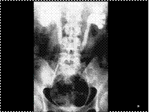
2. Retroperitoneal fibrosis. Bilateral retrograde pyelogram shows dilatation of the collecting systems and ureters to the level of the L4-L5 disk space with medial deviation of the ureters
Slide 22
Adrenal pseudotumor. A: An apparent mass is seen in the region of the left adrenal (arrow) on this computed tomography done without oral contrast. B: Following oral contrast administration, computed tomography shows the apparent mass was a gastric diverticulum. The normal left adrenal was seen more inferiorly
Slide 23
Pelvic lipomatosis. Medial deviation of the ureters associated with mild distal ureteral obstruction. The characteristic pear-shaped bladder is elevated by perivesical fat
Slide 24
3Urinary tract schistosomiasis. The characteristic curvilinear calcification in the bladder wall is seen on the precontrast radiograph of an IVU series.
Slide 25
Aortocaval fistula.
Slide 26
Von Hippel-Lindau (VHL). Multiple renal cysts present on contrasted CT are the most common renal lesions in this syndrome. The presence of pancreatic cysts (arrows) is also common in VHL. multiple enhancing renal masses (arrows) is highly suspicious for multifocal RCC in this setting
Slide 27
Adenomyosis. A: Sagittal T2-weighted fast spin-echo (FSE) (4500/108) magnetic resonance image shows enlargement of the uterus and a markedly thickened low signal junctional zone (arrowheads). Multiple foci of high T2 signal are present. B: Axial image using the same technique shows the high T2 foci to be endometrial glands extending into the adenomyoma (arrow). Bilateral high T2 signal ovarian cysts are present
Slide 28
Osteopathia striata. Anteroposterior radiograph of the right knee of a 14-year-old girl who had a history of trauma reveals, as an incidental finding, fine linear striations in the diaphysis and metaphysis of the distal femur and proximal tibia; the epiphyses, however, are spared.
Slide 29
Multiple bilateral oncocytomas. On contrasted CT (A, B), numerous enhancing renal masses enlarge the kidneys without distant metastases.
Slide 30
1.Medullary sponge kidney. (A) Excretory urogram demonstrates multiple small, smoothly rounded calculi occurring in clusters in the papillary tips of multiple renal pyramids. (B) In another patient, the ectatic tubules appear as fine linear striations of contrast producing the characteristic brush border pattern.
Slide 31
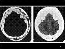
Cool phase of Paget disease:CT sections clearly demonstrate predominant involvement of the inner table with marked diminution of the diploic space (B) and thickening of the cranial vault
Slide 32
MAFUCCI SYNDROME (RIGHT, MULTIPLE ENCHONDROMAS AND ASSOCIATED SOFT TISSUE HEMANGIOMA WITH PHLEBOLITHS) , OLIERS DISEASE(LEFT)(MULTIPLE LARGE LOBULATED MASSES WITH ASSOCIATED BONE DEFORMITY)
Slide 33
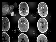
Fahr disease
Slide 34
GASTROJEJUNAL INTUSSUCCEPTION
Slide 35
Polyostotic fibrous dysplasia. A shepherd's crook deformity, seen here in the proximal femur in a 12-year-old boy with polyostotic fibrous dysplasia, is often the result of multiple pathologic fractures.
Slide 36
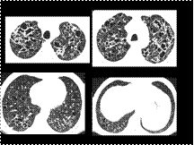
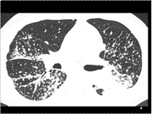
HRCT shows numerous, thick and thin-walled lung cysts. Some are very irregular in shape. Intervening lung parenchyma appears normal, and there is no evidence of fibrosis. Cysts are largest and most numerous in the lung apices (Figure 1). Cysts at the lung bases are smaller and less numerous. The left costophrenic angle appears spared Figure 4.
Diagnosis: Langerhans histiocytosis, presenting with lung cysts
DISCUSSION
Pulmonary histiocytosis, also known as Langerhans histiocytosis or eosinophilic granuloma of the lung, is an idiopathic disease characterized in its early stages by granulomatous nodules containing Langerhans histiocytes and eosinophils, which are primarily peribronchial in distribution.
In its later stages, the cellular granulomas are replaced by fibrosis and the formation of cysts.
Histiocytosis is an uncommon condition. The majority of patients with pulmonary histiocytosis are young or middle-aged adults presenting with nonspecific symptoms of cough and dyspnea. Up to 20% of patients present with pneumothorax. There is a slight male predominance, and over 90% of patients are smokers.
The HRCT findings of pulmonary histiocytosis closely mirror the gross pathologic appearances of this disease. In almost all patients, HRCT demonstrates cystic airspaces, which are usually less than 10 mm in diameter. On HRCT, the lung cysts have walls which range from being barely perceptible to being several millimeters in thickness. The presence of distinct walls allows differentiation of these cysts from areas of emphysema. Although many cysts appear round, they can also have bizarre shapes, being bilobed, clover-leaf shaped, or branching in appearance. An upper lobe predominance in the size and number of cysts is common, and the costophrenic angles are often spared.
In most patients, cysts are the only abnormality visible on HRCT, but in some, small nodules (usually less than 5 mm in diameter) are also present. In many patients with cysts or nodules the intervening lung parenchyma appears normal on HRCT, without evidence of fibrosis or septal thickening.
Slide 37
Lymphangiogram
Slide 38
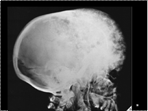
Lateral radiograph of the skull demonstrates predominant involvement of the frontal bones with a characteristic expansion of the outer table. The base of the skull, a frequent site of polyostotic fibrous dysplasia, is typically thickened, and the frontal and ethmoid sinuses are obliterated. The maxilla and mandible are also affected. This advanced stage of involvement of the skull and facial bones by polyostotic fibrous dysplasia is frequently termed leontiasis ossea.
Slide 39
Cavography Budd chiari syndrome
Slide 40
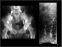
Sarcoidosis with perilymphatic nodules
Slide 41
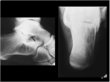
Simple bone cyst. Lateral radiograph of the hindfoot (A) and Harris-Beath view of the calcaneus (B) in a 32-year-old man show a simple bone cyst in the os calcis. Typically, bone cysts occurring at this site are located in the anterolateral aspect of the bone, as shown here.