Spotters A7

Slide 2
STRESS FRACTURE

Slide 3
Primary sclerosing cholangitis. Endoscopic retrograde cholangiopancreatography shows multifocal strictures (arrows) involving the intrahepatic and extrahepatic biliary ducts

Slide 4
GCT

Slide 5
Intrahepatic cholangiocarcinoma. Late hepatic arterial phase image (A) shows a large lobulated central hepatic mass with peripheral enhancement. A slightly more cephalad image (B) shows dilation of bile ducts peripheral to the mass and transient hepatic attenuation difference (THAD) with diffusely increased attenuation of the right hepatic lobe. A coronal 3D-volume rendered image (C) shows a satellite lesion (arrow) near the dome of the liver

Slide 6
Oesophageal duplication cyst

Slide 7
Epithelioid hemangioendothelioma. Unenhanced CT image (A) shows two peripheral hypoattenuating liver masses (M). The large right lobe mass represents coalescence of several smaller lesions that were present on prior examinations. Arterial (B), portal venous (C), and equilibrium phase (D) images demonstrate peripheral enhancement with gradual centripetal progression. Note the capsular retraction (arrows) associated with the masses and hypertrophy of the remaining normal hepatic parenchyma.

Slide 8
Uneven vascularity. In a patient with Macleod's syndrome a hypoplastic right main pulmonary artery leading to a reduction in vascular markings and a small right hemithorax are seen

Slide 9
Pancreatic trauma. Following direct blunt trauma to the abdomen the pancreas is seen to be fractured (arrow). There was disruption of the main pancreatic duct.

Slide 10
Usual interstitial pneumonia. HRCT abnormalities predominate in the posterior, subpleural regions of the lower lobes and comprise honeycombing and traction bronchiectasis within the abnormal lung.

Slide 11
EWING'S SARCOMA

Slide 12
Mirizzi syndrome. MRCP (A) shows a stricture of the lower common duct caused by a stone (arrow) lying in an expanded cystic duct on ERCP (B). Multiple gallbladder stones are also seen.

Slide 13
En face appearance of benign gastric ulcer. (A) Posterior wall ulcer is nearly filled with barium in this RPO projection. Thin regular radiating folds (best seen around the inferior border of the ulcer) are seen converging to the ulcer. (B) Unfilled benign ulcer crater is outlined by a ‘ring’ shadow. This ulcer is surrounded by a prominent ring of oedema – the lucent area around the crater.

Slide 14
Urinary tract schistosomiasis. The characteristic curvilinear calcification in the bladder wall is seen on the precontrast radiograph of an IVU series. Ureteric obstruction was seen following contrast injection (not shown).

Slide 15
Carcinoid tumour. A round, well-defined, intraluminal filling defect (arrow) is seen in the distal ileum of a patient who presented with symptoms of intermittent obstruction but without any manifestations of the carcinoid syndrome.
Carcinoid tumour. CT shows a carcinoid mass (arrow) with a characteristic stellate radiating pattern and thickening of the adjacent intestinal wall.

Slide 16
Pneumatosis coli. Multiple small gas-filled cysts are seen in association with the colon. There is also free peritoneal gas.

Slide 17
Fibromuscular dysplasia. (A) On this AP aortogram the pigtail catheter is positioned just above the renal arteries. There is fibromuscular disease involving the distal right renal artery (arrow) with an aneurysm (short arrow). (B) MR angiography demonstrates the same findings. (C) On a selective right anterior oblique (RAO) angiogram the characteristic saccular dilatations and the web-like stenoses are more clearly evident.

Slide 18
(A) Part of a panoramic radiograph showing a corticated radiolucency between the inferior alveolar canal and the lower border of the mandible due to the presence of a Stafne bone cavity. The 3D CT (B) shows the depression on the lingual aspect of the mandible.

Slide 19
Thyroid ophthalmopathy. (A) Axial and (B) coronal CT imaging. There is generalized enlargement of the bellies of all the extra-ocular muscles, proptosis and increased intraorbital fat.

Slide 20
Hepatocellular adenoma. A mass in segment VII of the liver is hyperintense on an in-phase T1-weighted gradient echo image (A) and shows marked signal loss on an opposed-phase image (B), indicating that it contains lipid. The mass is high in signal intensity on a T2-weighted image (C).

Slide 21
Right lower lobe collapse. (A) Frontal view of an example of right lower lobe collapse demonstrating a triangular density which does not obscure the right hemidiaphragm silhouette. (B) The lateral radiograph shows the typical features of increased density of the posterior costophrenic angle and loss of the silhouette of the right diaphragm posteriorly.

Slide 22
Scleroderma. (A,B) Two patients with scleroderma showing ground-glass opacification in association with traction bronchiectasis and a fine reticular pattern. The pattern of fibrosis is closest to that of non-specific interstitial pneumonia. Note the dilated oesophagus in both examples.

Slide 23
Lingular collapse. (A) Frontal view of isolated collapse of the lingular segments of the left upper lobe showing loss of clarity of the left heart border and a raised hemidiaphragm. (B) The similarity to a right middle lobe collapse can be appreciated on the lateral view.

Slide 24
Drusen. Axial CT. There are small foci of calcification at both optic nerve heads

Slide 25
Tetralogy of Fallot. Boot-shaped heart, small hila and pulmonary oligaemia.

Slide 26
Glomus jugulare tumour. An axial CT (A) demonstrates expansion of the right jugular foramen and bone destruction in the adjacent petrous bone by a mass that is markedly enhancing on axial T1W post-contrast images (B). The mass contains areas of flow voids, corresponding to the dilated tumour vessels seen on the right external carotid artery angiogram (C).

Slide 27
Pellegrini–Stieda lesion. Calcification/ossification is seen related to the superior portion of the medial femoral condyle (arrow).

Slide 28
Subfrontal meningioma. CT before (A) and after (B) IV contrast medium, and lateral projection of common carotid arteriogram (C). There is a large circumscribed mass in the anterior cranial fossa that is isodense to normal grey matter, contains foci of calcification centrally and enhances homogeneously. There is oedema in the white matter of both frontal lobes and posterior displacement and splaying of the frontal horns of the lateral ventricles. On the arteriogram (C) the mass is delineated by a tumour blush and there is posterior displacement of the anterior cerebral arteries (arrowhead), mirroring the mass effect seen on CT. The ophthalmic artery is enlarged as its ethmoidal branches supply the tumour (arrow).

Slide 29
pagets

Slide 30
Primary cerebral lymphoma. CT before (A) and after IV contrast medium (B). An irregular mass that is hyperdense to grey matter expands the splenium of the corpus callosum and extends into the left hemisphere. It is surrounded by extensive white matter oedema and enhances avidly with contrast.

Slide 31
Emphysematous pyelonephritis. Patient with diabetes mellitus and sepsis. The left renal collecting system and ureter are distended and gas filled. There are also multiple dense gallstones in the gallbladder.

Slide 32
Hiatal hernia, sliding type. A: A portion of the stomach (S), opacified with oral contrast, has passed through the diaphragm into the mediastinum. B: At a more caudal level, there is minimal widening of the esophageal hiatus (arrows).

Slide 33
Appendicitis. There is generalized ileus and an appendicolith in the right iliac fossa overlying the right side of the sacrum. At operation the appendix was gangrenous and perforated.

Slide 34
Obturator hernia causing small bowel obstruction. A: In this patient the hernia sac, which contains a small bowel loop (arrow), passes between the external (arrowhead) and internal (open arrow) obturator muscles, a less common form of obturator hernia than that seen in Figure 16-6.

Slide 35
Scapholunate dissociation. PA view of the wrist in ulnar deviation (A) shows abnormal widening of the scapholunate distance (greater than 4 mm), consistent with disruption of the scapholunate ligament. View in radial deviation (B) demonstrates no significant abnormality; the widening is not apparent.

Slide 36
Wilson's Disease
Wilson's disease is a rare autosomal recessive, inborn error of copper metabolism characterized by abnormal accumulation of copper in various tissues, mainly in the brain and liver.
Parkinsonism, tremor, ataxia, and dystonia are common clinical features. Presence of Kayser-Fleischer ring in the cornal limbus is diagnostic.
Key imaging features: Bilateral, symmetric increased signal intensity on T2WI is most often seen involving the putamina. Similar bilateral abnormal signal intensity can be seen involving the caudate nuclei, thalami, red nuclei, and dentate nuclei. Both pyramidal and extrapyramidal white matter tracts can be involved. ""Face of giant panda"" and ""bright claustral"" signs are often seen.
DDx: Hypoxic-ischemic encephalopathy
Treatment: D-penicillamine, Vit E, Pyridoxine, Low Cu diet
Slide 37
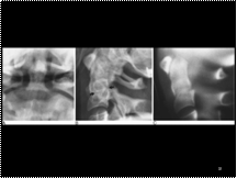
Low (Type III) dens fracture. (A) AP open-mouth view demonstrates lateral tilting of the dens. No fracture line is apparent. This is frequently the case in this type of fracture. (B) Lateral view demonstrates disruption of the ring of C2 (arrows). There is slight anterior tilting of the long axis of the dens. The dens is usually either in neutral or posterior angulation relative to the body of C2 in lateral projection. (C) A lateral tomogram clearly demonstrates the fracture line and the tilting of the dens.

Slide 38
HRCT of the ‘crazy paving’ pattern in alveolar proteinosis: patchy but geographical ground-glass opacification is seen and there are numerous thickened interlobular septa in areas of ground-glass opacification.

Slide 39
CT arthrography: bucket handle medial meniscal tear—correlation with MR and CT. A: Coronal fat-suppressed intermediate-weighted spin-echo image and (B) coronal reformation obtained after spiral CT arthrography of the left knee of a 27-year-old man shows a tear of the body of the medial meniscus with a meniscal fragment flipped in the intercondylar notch. The lesion was confirmed at arthroscopy

Slide 40
ENCEHALOCELE/CH
Slide 41
Moyamoya Disease
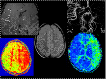
Moyamoya disease is a rare vascular disorder characterized by progressive, idiopathic narrowing of the supraclinoid segment of the internal carotid artery (ICA), with secondary collateralization.
Seen more often in children and young females, it presents with developmental delay, poor feeding, TIAs, and alternating hemiplegia.
Key imaging features: FLAIR: Bright sulci, asymmetric presence of prominent VR spaces, less often asymmetric ischemic foci. MRA: Narrowing of distal ICA and proximal circle of Willis vessels, and collateralization. ""Puff or spiral of smoke"" on angiography. Perfusion imaging: Hypoperfused areas which can guide further management.
DDx: Subarachnoid hemorrhage, meningeal carcinomatosis, meningitis, high inspired oxygen, carotid dissection.
Treatment: Medical and surgical: encephalo-duro-arterio-synangiosis (EDAS). Surgery helps but does not prevent the progression of disease
Slide 42
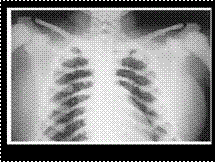
OSTEOPETROSIS
Slide 43
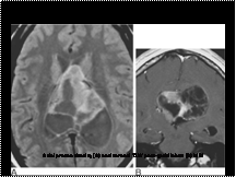
Central neurocytoma. Axial proton density (A) and coronal T1W post-gadolinium (B) MRI. A partly cystic, multi-septated, enhancing mass, which is related to the septum pellucidum, fills the bodies of both lateral ventricles and causes hydrocephalus with dilatation of the left temporal horn
Slide 44
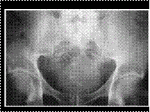
SACRAL CHORDOMA. AP Pelvis. Observe the destructive lesion in the distal surface of the sacrum.
Slide 45
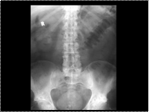
UC
Slide 46
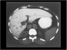
Haemochromatosis. Unenhanced CT section through the liver of a patient with haemochromatosis showing diffuse increased attenuation of the liver compared with the spleen
Slide 47
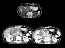
Pseudoaneurysm of gastroduodenal artery
Slide 48
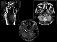
Persistent Hypoglossal Artery
The persistent hypoglossal artery (PHA) is the second most common persistent carotid-basilar anastomosis after the trigeminal artery.
PHA originates from the posterior side of the cervical part of the internal carotid artery at the level of C1-C2 and enters the skull through an enlarged hypoglossal canal; it ends in the basilar artery.
When present, PHA is often the exclusive feeder of the posterior circulation and is associated with hypoplasia of the vertebral arteries.
This vascular variant can be associated with hypoglossal nerve palsy, basilar artery aneurysm, and cranio-cervical junction bone malformations.
Temporary clamping of the hypoglossal artery during CEA carries greater ischemic risk.
Differential diagnosis: type I proatlantal artery enters the skull through the foramen magnum.
Slide 49
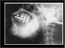
FIBROUS DYSPLASIA: CHERUBISM
Slide 50
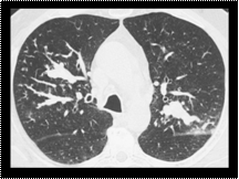
Allergic bronchopulmonary aspergillosis. HRCT of the upper lobes. Mucoid impactions are present within segmental and subsegmental dilated bronchi in the upper lobes. Small centrilobular linear branching opacities are seen in the periphery of the right upper lobe.
Slide 51
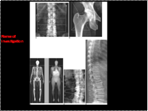
Quantitative assessment of the skeleton—DXA. Dual energy X-ray absorptiometry (DXA) provides ‘areal’ BMD (g cm-2) and is currently the ‘gold standard’ method for diagnosis of osteoporosis by bone densitometry (WHO definition T score -2.5 or below) in (A) PA lumbar spine (L1–4) or (B) hip (femoral neck or total). (A) Osteophytes at L2/3 and L3/4 will cause false elevation of bone mineral density (BMD) in this anatomical site, especially in the elderly. DXA of the whole body can provide information on (C) total and regional BMD and (D) body composition (fat and muscle mass). (E) Vertebral assessment can be made from lateral images (single and dual energy; latter superior for imaging thoracic region) obtained on fan beam DXA systems at about 1/100th of the dose of conventional radiography (fractures present at L2).

