Spotters A9

Slide 2
(B) Rickets in the wrist of a child. There is increase in the width of the growth plate and cupping and splaying of the metaphysis, which is poorly mineralized. These radiographic features can occur as a result of vitamin D deficiency (lack of active metabolite 1,25(OH)2D3) from nutritional lack (dietary or lack of sunlight), malabsorption, liver disease or chronic renal failure, hypophosphataemia, hypophosphatasia, acidaemia and low calcium intake.

Slide 3
BASILAR IMPRESSION WITH OCCIPITALIZATION

Slide 4
Intrathoracic thyroid mass on (A) AP and (B) lateral radiographs. This benign multinodular goitre is predominantly posterior to the trachea with components to either side, resulting in forward displacement and narrowing of the trachea.

Slide 5
Spinal schwannomas

Slide 6
Synovial osteochondromatosis. Lateral radiograph showing the fine cartilage calcifications of primary synovial osteochondromatosis.

Slide 8
Germinoma of the suprasellar region

Slide 9
Right middle lobe collapse. An example showing a triangular-shaped density adjacent to the right heart border

Slide 10
Aseptic meningitis

Slide 11
DAI

Slide 12
High jugular bulb.

Slide 13
Panlobular emphysema in a patient with ?1-antitrypsin deficiency. HRCT of the lung bases showing the presence of large areas of decreased lung attenuation with a paucity of pulmonary vessels, more marked in the lower lobes. Long lines are visible within the remaining parenchyma of lung bases

Slide 14
Teratoma in a young man undergoing an immigration chest radiograph. (A) There are no specific features on the plain radiograph to indicate the nature of the mass. (B) CT demonstrates that the opacity visible on the chest radiograph is well defined and contains soft tissue and fat densities.

Slide 15
Neural tube defects. (A) Lemon-shaped skull associated with a neural tube defect. The cisterna magna is effaced due to an Arnold–Chiari malformation. (B) In the same fetus the coronal view of the lumbar shows splaying of the posterior ossification centres in a fetus with an open neural tube defect.

Slide 16
Chondromyxoid fibroma. Lateral radiograph of the proximal tibia showing expansion of the anterior cortex

Slide 17
Thornwaldt's cys

Slide 18
Pyeloureteritis cystica. A staghorn calculus occupies much of the collecting system.

Slide 19
Carcinoma of the Esophagus: Varicoid Pattern1

Slide 20
OSTEOPOIKILOSIS

Slide 21
Ganglioglioma of the right temporal lobe

Slide 22
CANAVAN

Slide 23
ACHONDROPLASIA

Slide 24
Meningioma arising from the posterior aspect of the left temporal bone

Slide 25
Fibrosing mesenteritis. Enhanced CT in a patient who presented with fever of unknown origin demonstrates a fibrofatty mesenteric mass with irregular borders surrounding mesenteric vessels. Strands of soft-tissue density are seen radiating from the mass to the adjacent mesenteric fat.
Fibrosing mesenteritis: CT appearances. Enhanced abdominal CT demonstrating a large, ill-defined, soft-tissue mesenteric mass with extensive calcification. Note retraction and thickening of the adjacent bowel loops.

Slide 26
Pneumatosis coli with numerous gas cysts in the wall of the colon.

Slide 27
Hypertrophic gastritis in a patient with a recently healed lesser curvature gastric ulcer. This characteristic enlargement and prominence of the areae gastricae can be correlated with an increased incidence of gastric hypersecretion and PUD.

Slide 28
Gastric bezoar. Large mass in stomach – solidified retained ingested fibrous material mixed with air in the patient after a Billroth II anastomosis

Slide 29
Lipoma. (A) On mammography, a lipoma may be seen as a well-defined mass of fat density, contained within a thin capsule. (B) On ultrasound, a well-defined hyperechoic lesion characteristic of a lipoma is seen.

Slide 30
Hepatocellular carcinoma. Contrast-enhanced CT images (A, B) show a large heterogeneously enhancing mass in the right hepatic lobe. Internal septations and areas of degeneration give the mass a “mosaic†pattern, which is also demonstrated on a coronal volume-rendered image (C). The very low attenuation areas in the inferior portion of the mass are areas of fatty degeneration.

Slide 31
Medullary sponge kidney. (A) Plain radiograph demonstrates multiple foci of calcification which are seen to lie in the renal medulla after injection of contrast medium (B).

Slide 32
Craniopharyngioma in a child

Slide 33
TOF

Slide 34
Ependymoma of the fourth ventricle

Slide 35
Salter–Harris Type I epiphyseal separation of the proximal tibia.

Slide 36
Ovarian dermoid, intrauterine pregnancy. T2-weighted sagittal (A) and T1-weighted axial (B) MRI. The sagittal image demonstrates a fetus (F) in vertex position. Situated posterior to the uterus in the cul-de-sac is a hyperintense cystic and solid, rounded mass (M). A portion of the mass is high signal on T1-weighted image and falls in signal intensity following a fat saturation pulse (arrow B). This is consistent with fat, allowing the diagnosis of dermoids to be made with confidence.
Slide 37
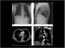
PTE

Slide 38
Myelofibrosis. (A) Lateral radiograph of the lumbar spine showing diffuse vertebral body sclerosis. (B) Coronal T1-weighted spin-echo MRI showing patchy reduction of marrow signal intensity in the pelvic bones and proximal femora.
Slide 39
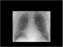
Right middle lobe collapse. An example showing a triangular-shaped density adjacent to the right heart border

Slide 40
Ileocolic intussusception and subsequent infarcted closed-loop obstruction. 44-year-old woman with metastatic melanoma. A: There is an intussusception in the ascending colon (asterisk). It contains fat and soft tissue. B: Slightly more caudal image demonstrates the intussusceptum to contain terminal ileum (arrow) and mesenteric fat with vessels. C: More cranial image reveals soft tissue mass (asterisk) as the lead point. Melanoma metastasis was identified at surgery. This was resected and a primary anastomosis performed. Patient presented 9 months later with abdominal pain and vomiting. D: CT image demonstrates a closed-loop obstruction with a whirled appearance of loops of dilated, fluid-filled small bowel. There is mild bowel wall thickening with inflammatory changes in the mesentery and ascites. Collapsed distal small bowel lies adjacent to anastomotic line (arrow) from prior metastasis resection. Second surgery confirmed an internal hernia from a postsurgical rent in the mesentery with bowel infarction.

Slide 41
Septo-optic dysplasia in a child with HESX1 gene mutation. (A) The septum pellucidum is absent and the frontal horns have a typical box-like configuration. (B) The posterior pituitary gland is ectopic (arrow). (C,D) The right optic nerve is small (arrows).
Slide 42
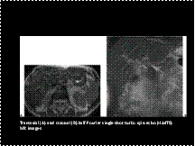
Biliary hamartomas in two patients. Transaxial (A) and coronal (B) half-Fourier single shot turbo spin echo (HASTE) MR images show multiple small hyperintense lesions scattered throughout the liver in each patient.
Slide 43
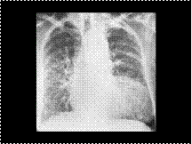
Bilateral lymphangitic carcinomatosis showing bilateral thickened septal lines together with widespread nodulation of the lungs. The primary tumour in this 71 year old woman was presumed to be a bronchial carcinoma (a diagnosis based on sputum cytology).
Slide 44
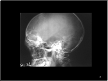
FRONTAL SINUS MUCOCELE
Slide 45
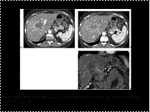
Intrahepatic cholangiocarcinoma. Late hepatic arterial phase image (A) shows a large lobulated central hepatic mass with peripheral enhancement. A slightly more cephalad image (B) shows dilation of bile ducts peripheral to the mass and transient hepatic attenuation difference (THAD) with diffusely increased attenuation of the right hepatic lobe. A coronal 3D-volume rendered image (C) shows a satellite lesion (arrow) near the dome of the liver
Slide 46
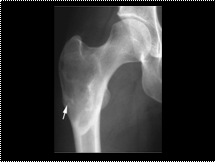
Fibrous dysplasia. AP radiograph of the proximal femur showing a well-defined expanded lesion with typical ground-glass matrix mineralization and a thick, sclerotic margin (rind sign). A stress fracture is present in the lateral cortex (arrow).
Slide 47
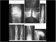
Hyperparathyroidism. In primary hyperparathyroidism there may be chondrocalcinosis (calcification of cartilage) as illustrated in (A) the knee and (B) the symphysis pubis. Other sites where this may be present are the triradiate ligament of the wrist and the large joints (hip and shoulder). Because of increased osteoclastic activity (C), subperiosteal erosions may be present and are most likely to be evident radiographically along the radial side of the middle phalanges of the 2nd and 3rd fingers. Acro-osteolysis is also present. As there is calcification of the digital artery (metastatic calcification), this is secondary hyperparathyroidism related to chronic renal failure (phosphate retention). Also related to increased osteoclastic resorption there can be (D) cortical ‘tunnelling’ (areas of resorption within the bone cortex) as in the proximal phalanges. (E) Bone cysts (brown tumours, osteitis fibrosa cystica) can occur in any site, seen here in the distal tibia. These cysts are less commonly seen now, as the diagnosis may come to attention in mild or asymptomatic patients who are found to have hypercalcaemia on routine blood testing. In hyperparathyroidism secondary to chronic renal disease there is phosphate retention, increase in the phosphate × calcium product and precipitation on amorphous calcium phosphate in the vessels and in soft tissues, usually around large joints, as seen (F) in the left shoulder.
Slide 48
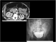
CT of intraperitoneal bladder rupture. (A) CT image obtained through the upper abdomen in the arterial phase shows low-attenuation fluid (nonopacified urine) in the perihepatic space (arrow) and left paracolic gutter (curved arrow). (B) Cystogram shows contrast medium over the dome of the bladder in a ‘molar tooth’-shaped configuration and ascending up the right paracolic space.
Slide 49
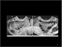
Polycystic ovaries. Sagittal (A) and transverse (B) transvaginal ultrasound of the left ovary depicting multiple subcentimetre peripherally placed follicles in enlarged ovaries with echogenic central stroma
Polycystic ovarian disease (Stein–Leventhal syndrome)
Polycystic ovarian disease is characterized by bilaterally enlarged polycystic ovaries, secondary amenorrhoea or oligomenorrhoea and infertility. About 50% of patients are hirsute and many are obese. Many cases of female infertility secondary to failure of ovulation are due to polycystic ovarian disease. The classic appearance on ultrasound is enlarged ovaries with echogenic central stroma and greater than 10 peripherally placed cysts less than 9 mm in diameter ( Fig 54.22 ).
Slide 50
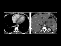
Azygos continuation of inferior vena cava
Slide 51
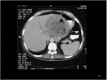
EMPHY PANCREATITIS


