Spotters A12

Slide 2
SIGMOID VOLVULUS

Slide 3
WORMS

Slide 4
RETROCAVAL URETER

Slide 5
EHPVO

Slide 6
ACUTE EPIGLOTITIS

Slide 7
PANCRETIC #

Slide 8
R STAGHORN CALCULUS

Slide 10
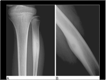
Melorheostosis (two patients). There is dense irregular cortical bone: this is sometimes described as the flowing candle wax appearance. One patient demonstrates a ray' distribution (asymmetrical changes).

Slide 11
RECTAL CA

Slide 12
Fibrous dysplasia. (A) Patchy sclerosis affecting the vault and the base of the skull. (B) Radiolucent areas in the femoral necks and pelvic bones. Bilateral coxa vara. This is an early shepherd's crook deformity. Widening of the upper femoral epiphyseal plates, due to associated hypophosphataemic rickets. (C) Multiple radiolucent areas causing expansion. The margins are sclerotic and extend over several millimetres (a 'rind' appearance). Scalloping of the cortex. (D) The radiolucent areas predominantly affect the radius in a ray distribution. (E) The metacarpals and phalanges of the second to fifth fingers show expansion and deformity. Multiple radiolucent clearly defined lesions with relative sparing of the thumb and carpus.

Slide 13
Fournier gangrene

Slide 14
OI

Slide 15
FUNGAL BALL

Slide 17
BUDD CHAIRI

Slide 18
RML COLLAPSE

Slide 19
ARCUATE UTERUS

Slide 20
POLYCYSTIC DS

Slide 21
Cerebral AVM on DSA. (A) Arterial and (B) venous phase of DSA in a patient with a cerebral AVM. The AVM is fed by branches of the anterior and middle cerebral arteries and the venous drainage is predominantly superficial into the superior sagittal and transverse sinuses.

Slide 22
L CDH

Slide 23
Pericardial effusion

Slide 24
Neuronal heterotopia. Coronal T1-weighted images from a volumetric acquisition; slice thickness is 1.5 mm. (A) Subependymal (nodular) heterotopia (arrowhead), (B) laminar heterotopia (arrowheads)

Slide 25
BRODIES ABCESS

Slide 26
KOHLERS DS

Slide 27
Ependymoma of the filum terminate and conus medullaris. Sagittal T2W (A) and (B) T1W post gadolinium-enhanced MRIs of the lumbar spine showing an expansile enhancing intraspinal mass and central signal change in the spinal cord above

Slide 28
LEPROSY

Slide 29
HYDATID DS

Slide 31
Apert's syndrome. (A,B) 3D CT surface-shaded display shows the wide open defect of the sagittal suture and brachycaphaly with bicoronal synostosis typical of Apert's syndrome. The coronal sutures appear fused and are ridged. (C,D) Plain radiographs of the hands show the ‘mitten hand’ appearance with syndactyly and shortened metacarpals

Slide 32
Optic nerve meningioma. (A) Axial T2, (B) axial T1 MRI with gadolinium and fat suppression. There is a mass at the right orbital apex, closely applied to the optic nerve but seen separate to it. On the contrast-enhanced image, ‘tram-track’ enhancement along the nerve can be seen

Slide 33
GCT

Slide 34
Esthesioneuroblasto-ma. (A) Contrast-enhanced sagittal T1-weighted MRI of mass based on cribriform plate. Note the extension inferiorly into the nose and superiorly into the brain. (B) Coronal CT shows the bony destruction of the cribriform plate and a soft tissue mass in the ethmoid. The differential diagnosis would include an ethmoid carcinoma

Slide 35
Congenital cystic adenomatoid malformation of lung (CCAM). A 4 year old child presented with acute dyspnoea. Chest radiograph shows a cystic lesion in the left lower lobe, confirmed histologically as a CCAM type 1

Slide 36
Low (Type III) dens fracture. (A) AP open-mouth view demonstrates lateral tilting of the dens. No fracture line is apparent. This is frequently the case in this type of fracture. (B) Lateral view demonstrates disruption of the ring of C2 (arrows). There is slight anterior tilting of the long axis of the dens. The dens is usually either in neutral or posterior angulation relative to the body of C2 in lateral projection. (C) A lateral tomogram clearly demonstrates the fracture line and the tilting of the dens

Slide 37
Osteoid osteoma. (A) Intracortical osteoid osteoma of the left fibular shaft: typical oval radiolucent area, representing the nidus, with calcification inside and reactive bone sclerosis. (B) Intracortical osteoid osteoma of the left femoral shaft without calcification in the radiolucent area and perifocal osteosclerosis

Slide 38
Dandy–Walker malformation. (A,B) The fourth ventricle opens into a large posterior fossa cyst. There is associated hydrocephalus. (C) The cerebellum is hypoplastic and a thin rim of cerebellar tissue is seen forming the wall of the posterior fossa cyst (arrow). The vein of Galen, straight sinus, and venous confluence are elevated above the level of the lambdoid suture

Slide 39
MRI, adenomyosis. Sagittal (A) and axial (B) T2-weighted MR images through the pelvis demonstrate focal junctional zone widening and multiple punctate high signal intensity foci with the areas of thickening (*, A, B) characteristic of focal adenomyosis. Sagittal (C) and axial (D) T2-weighted MR images in a different patient demonstrate widening of the entire junctional zone (*, C, D) which contains multiple foci of high signal intensity that represent endometrial rests. Appearances are typical of diffuse adenomyosis

Slide 40
CYSTIC FIBROSIS

Slide 41
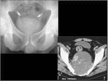
Sacral chordoma. AP radiograph of the sacrum shows a central, lytic destructive lesion (arrows).
CT shows a predominantly lytic mass with small foci of calcification


