Spotters B2
Slide 2
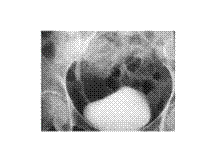
CHPS
Slide 3
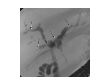
Primary sclerosing cholangitis. Endoscopic retrograde cholangiopancreatography shows multifocal strictures (arrows) involving the intrahepatic and extrahepatic biliary ducts.
Slide 4
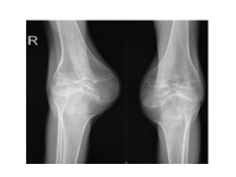
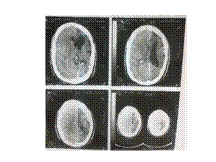
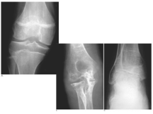
JRA
Slide 6
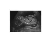
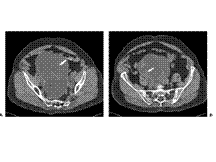
Fetal hydrops. Longitudinal view of a fetus with skin oedema, ascites and a hydrothorax
Slide 7
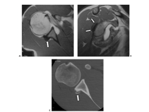
Bennett lesion. A: Axial T1-weighted MR arthrogram. There is globular low signal intensity suggestive of calcification along the posterior capsular insertion onto the glenoid (arrow). The subscapularis tendon is partially torn. B: Sagittal T1-weighted MR arthrogram image shows the extent of the posterior capsular low-signal abnormality (arrows). C: Transverse CT image confirms the presence of calcification of the posterior capsular insertion.
Slide 8
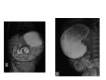
Mirizzi syndrome. MRCP (A) shows a stricture of the lower common duct caused by a stone (arrow) lying in an expanded cystic duct on ERCP (B). Multiple gallbladder stones are also seen.
Slide 9
Thyroglossal cyst
Slide 10
SCURVY
Slide 11
Fibrosing mesenteritis. Enhanced CT in a patient who presented with fever of unknown origin demonstrates a fibrofatty mesenteric mass with irregular borders surrounding mesenteric vessels. Strands of soft-tissue density are seen radiating from the mass to the adjacent mesenteric fat. Fibrosing mesenteritis: CT appearances. Enhanced abdominal CT demonstrating a large, ill-defined, soft-tissue mesenteric mass with extensive calcification. Note retraction and thickening of the adjacent bowel loops.
Slide 12
A focal tear is seen within the TFCC (arrow) on (A) coronal T1 and (B) GE images
Slide 13
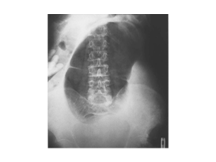
Caecal volvulus. The dilated caecum lies with its pole under the left hemidiaphragm. In spite of the dilatation the haustra are preserved. There is no dilated large bowel elsewhere in the abdomen. The small bowel is fluid filled in this case.
Slide 14
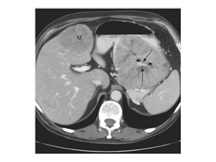
Gastric adenocarcinoma mimicking a GIST. There is a large heterogeneous mass in the body of the stomach. It is round and contains an ulcer (arrows). A large metastasis lies in the medial segment of the liver (M). It has similar enhancement characteristics as the primary tumor. Diagnosis of GIST was entertained because of the exophytic, ulcerated appearance, but endoscopic biopsy confirmed it to be a well-differentiated adenocarcinoma.
Slide 15
Ureteric obstruction. (A) IVU demonstrates high-grade obstruction in the right side of a horseshoe kidney
Slide 16
Colonic lipoma. There is a 2-cm fat-density mass in the proximal transverse colon (arrow). It is well defined and nonobstructing. This was an incidental finding. Lipomas can be distinguished from fat-density bowel contents by their notable lack of internal gas.
Slide 17
HEMANGIOMA
Slide 18
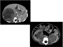
Cystadenoma. A and B: Axial contrast-enhanced computed tomography images show a large fluid density cystic mass with multiple predominantly thin septations (arrows) and no associated ascites.
Slide 19
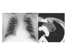
Dense pericardial calcification demonstrated on (A,B) chest radiograph (arrows) and (C) CT. There are bilateral pleural effusions in this patient with constrictive calcific pericarditis due to previous tuberculosis.
Slide 21
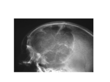
Thalassaemia. Lateral radiograph of the skull showing gross expansion of the diploë and loss of definition of the outer table with sparing of the occipital bone. A gross ‘hair-on-end’ appearance is shown
Slide 22
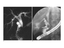
Grade III urethral injury. Retrograde urethrogram performed with a Foley catheter in the distal penile meatus reveals extravasation of contrast material from the posterior urethra extending above and below the level of the urogenital diaphragm. Some venous uptake is also seen.
Slide 23
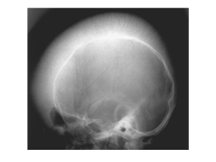
Atretic parietal encephalocele
Slide 24
Emphysematous cholecystitis. (A) CT – Intraluminal gas; (B) US – intraluminal gas appears as a bright curvilinear echogenic band (arrow) with ‘dirty’ shadowing.
Slide 25
Haemophilia. (A) Antero-posterior radiograph of the knee showing epiphyseal overgrowth and enlargement of the intercondylar notch. (B) Antero-posterior radiograph of the elbow showing erosion of the radial notch of the ulna. (C) Antero-posterior radiograph of the ankle showing medial tibiotalar slant and secondary osteoarthritis.
Slide 26
Breast implant
Slide 27
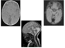
TAPVC
Slide 28
Biliary cystadenoma in a 49-year-old woman. Fast spin-echo T2-weighted MR image shows a multilocular, septated mass (arrows) in segment 7 of the liver, with high signal intensity within the cystadenoma. Corresponding portal-venous-phase gadolinium-enhanced T1-weighted MR image shows enhancement of the capsule and septa
Slide 29
pseudomyxoma peritonei
Slide 30
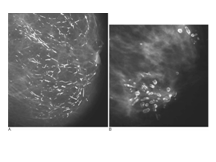
Duct ectasia. (A) Broken needle appearance, typical of duct ectasia. (B) Sometimes thicker, more localized calcifications can be seen, giving a ‘lead-pipe’ appearance
Slide 31
Transitional cell tumour seen on an IVU as a filling defect in the distal ureter.
Slide 32
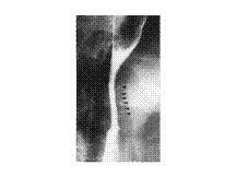
Fibrous dysplasia in a rib; chest radiograph detail of the left lung. Compared with the other ribs the ninth rib shows an increase in density and is slightly broadened
Slide 33
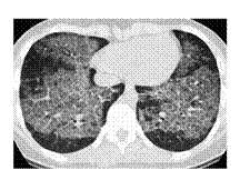
Pulmonary alveolar proteinosis, 15-year-old girl with shortness of breath. High-resolution computed tomography through the lung bases shows extensive ground-glass opacity and interstitial thickening, creating a “crazy paving†appearance

Slide 34
Pneumoperitoneum resulting from perforation of a duodenal ulcer. Erect chest radiograph. Typical free gas between the liver and the right hemidiaphragm. Note also the small triangular collection between the loops of the splenic flexure of the colon, beneath the left hemidiaphragm
Slide 35
Barium swallow demonstrating the typical appearances of oesophageal intramural pseudodiverticulosis. The small flask-shaped pits of contrast (arrowheads) represent dilated mucous glands and are associated with a stricture at the level of the aortic knuckle
Slide 36
Urinary tract schistosomiasis. The characteristic curvilinear calcification in the bladder wall is seen on the precontrast radiograph of an IVU series. Ureteric obstruction was seen following contrast injection (not shown)
Slide 37
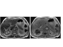
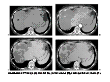
Epithelioid hemangioendothelioma. Unenhanced CT image (A) shows two peripheral hypoattenuating liver masses (M). The large right lobe mass represents coalescence of several smaller lesions that were present on prior examinations. Arterial (B), portal venous (C), and equilibrium phase (D) images demonstrate peripheral enhancement with gradual centripetal progression. Note the capsular retraction (arrows) associated with the masses and hypertrophy of the remaining normal hepatic parenchyma
Slide 38
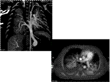
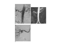
Fibromuscular dysplasia. (A) On this AP aortogram the pigtail catheter is positioned just above the renal arteries. There is fibromuscular disease involving the distal right renal artery (arrow) with an aneurysm (short arrow). (B) MR angiography demonstrates the same findings. (C) On a selective right anterior oblique (RAO) angiogram the characteristic saccular dilatations and the web-like stenoses are more clearly evident
Slide 39
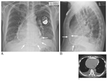
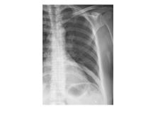
Teratoma in a young man undergoing an immigration chest radiograph. (A) There are no specific features on the plain radiograph to indicate the nature of the mass. (B) CT demonstrates that the opacity visible on the chest radiograph is well defined and contains soft tissue and fat densities
Slide 40
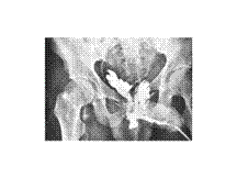
WILMS
Slide 41
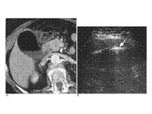
MULTIPLE ENCHONDROMAS INVOLVING
ENTIRE FEMUR ALONG
WITH SOFT TISSUE HAEMANGIOMAS