Spotters B6

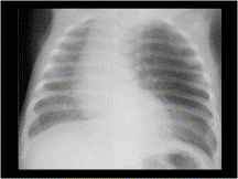
Slide 2
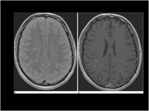
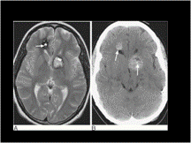
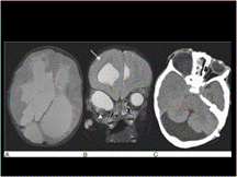
CLE
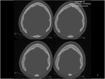
Slide 3
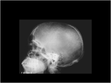
Giant parietal foramen
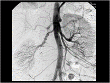
Slide 4
FMD
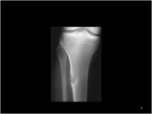
Slide 5
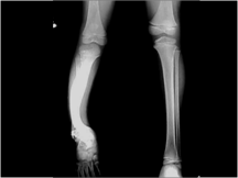
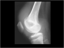
Figure 47.35 AP radiograph of the proximal tibia showing a diaphyseal fibrous cortical defect. Note the ground-glass appearance of the matrix
Slide 6
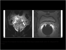
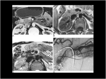
Figure 20-33 Advanced prostate cancer. A: Coronal T2-weighted fast spin-echo (FSE) image (4000/108) of the prostate obtained using an endorectal coil shows diffuse abnormal low signal throughout the peripheral zone. These are multiple sites of adenocarcinoma. The ejaculatory ducts are also thickened (arrows). B: Axial T2-weighted FSE image using the same pulse sequence as in A shows thickening of the normally gracile seminal vesicle septa indicating tumor invasion (arrows)
Slide 7
Neurofibromatosis
Slide 8
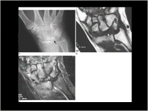
Figure 50.37 Kienböck's disease. (A) The lunate is collapsed and sclerotic indicating avascular necrosis (arrow). (B) T1-weighted MR image. The normal fatty marrow is replaced by low signal material indicating that the bone is avascular (arrow). (C) Fat-saturated FSE T2-weighted sequence shows a paucity of inflammatory change
Slide 9
Osteosarcoma
Slide 10
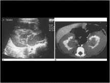
NEC
Slide 11
Median arcuate ligament syndrome
Slide 12
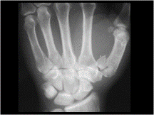
Rolando’s #
Slide 13
Zenk divert
Slide 14
Multiple sclerosis. (A) Axial proton density and (B) gadolinium-enhanced T1W images show multiple lesions in the periventricular white matter, two of which are in the acute phase and enhance.
Tumefactive MS. (A) Axial T2W fast spin-echo and (B) gadolinium-enhanced T1W spin-echo images show a large MS plaque with considerable associated oedema in the right frontal lobe. Note the smaller periventricular lesions on the T2W image, and the ‘open ring’ of contrast enhancement.
Slide 15
PSEUDOANEURYSM
Slide 16
5th nerve schwanoma
Slide 17
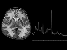
canavan
Slide 18
Ica aneurysm
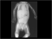
Slide 19
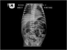
volvolus
Slide 20
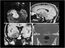
SEMILOBAR HOLOPRESENCEPHALY

Slide 21
L atrial enlargement
Slide 22
LYMPHANGIOMA OF NECK
Slide 23
Cavernous haemangioma. (A) T2-weighted axial image showing typical mixed signal intensity lesions. High signal is due to methaemoglobin and the low signal intensity rim of haemosiderin indicates an old haemorrhage. The ‘popcorn’ appearance of the larger lesion is typical of a ‘cavernoma’. Note the blood–fluid level in the smaller lesion (arrow). (B) Unenhanced CT of the same patients shows the lesions to be predominantly high density with tiny foci of calcification (arrows)
Slide 24
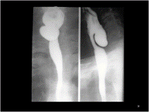
PORTAL CAVERNOMA
Slide 25
Figure 24-26 Extralobar pulmonary sequestration, 6-month-old boy with a left paraspinal mass on chest radiographs. A: Computed tomography (CT) scan shows the anomalous vein (arrow) arising from the sequestered lung (S) and crossing the midline to the right hemithorax. B: More caudal CT image demonstrates the anomalous arterial vessel (white arrow) arising from the celiac artery (black arrow). A, aorta
Slide 26
ASPEGILOSIS
Slide 27
Hyperparathyroidism
Slide 28
Figure 66.29 Nephroblastomatosis. (A) US demonstrating uniformly hypoechoic lesions surrounding the periphery of an enlarged kidney in a diffuse type of nephroblastomatosis. (B) Delayed post-contrast CT sections demonstrate these lesions, typically showing reduced vascularity in a fairly homogeneous pattern. Dense contrast medium is seen within the normal renal parenchyma.
The differential diagnosis of diffuse nephroblastomatosis on US mainly includes renal lymphoma or leukaemia
Slide 29
Figure 67.10 Chondrodysplasia punctata. Stippled or punctate calcification in the region of the epiphyses
Slide 30
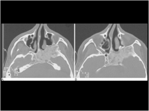
Figure 62.13 Fibro-osseous lesions. (A) The milky texture of bone typical of fibrous dysplasia is seen in this CT. (B) Fibrous dysplasia is a lesion which expands bone—note the compression of the pterygopalatine fissure
Slide 31
Figure 74.27 Haemophilia. Lateral radiograph of the knee shows extensive, hyperdense synovitis
Slide 32
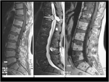
Figure 74.18 Multiple myeloma. (A) Sagittal T1-weighted spin-echo and (B) fat-suppressed T2-weighted fast spin-echo MRI showing pathological collapse of T11 with cord compression and multiple smaller areas of focal marrow involvement. (C) Sagittal T1-weighted spin-echo MRI showing the variegated pattern of marrow involvement
Slide 33
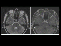
Figure 61.10 Optic nerve meningioma. (A) Axial T2, (B) axial T1 MRI with gadolinium and fat suppression. There is a mass at the right orbital apex, closely applied to the optic nerve but seen separate to it. On the contrast-enhanced image, ‘tram-track’ enhancement along the nerve can be seen
Slide 34
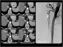
AAA
Slide 35
Figure 71.17 Insulinoma. (A) Contrast enhanced spiral CT demonstrating a small brightly enhancing insulinoma (curved arrow) in the head of the pancreas. (B) Axial T1-weighted spin echo MR image and (C) T2-weighted fast spin echo image show the corresponding lesion (arrow) on MRI of low signal intensity on the T1- and high signal intensity on the T2-weighted images. (D) A coeliac arteriogram in the same patient shows the vascular blush of the insulinoma (arrow)
Slide 36
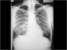
RETICULO NODULAR
GRANULOMATOUS - VIRAL- CHICKEN POX
T.B.
HISTO- OTHER FUNGAL
EOSINOPHILIA
SARCOID
OCCUPATIONAL - Anthraco, Berrylliosis
EMBOLIC - Fat embolism
IDIOPATHIC - Haman rich
MALIGNANCY - leukamia, lymphoma
Alveolar cell carcinoma
METASTASIS - Lympho hematogram - Thyroid
- Colon, pancreas
- prostate
- Breast in female
Slide 37
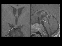
Figure 70.52 Hypothalamic hamartoma. Coronal T1 and sagittal T1-weighted post-contrast MRI shows a nonenhancing lesion arising from the floor of the third ventricle posterior to the pituitary infundibulum and projecting inferiorly into the suprasellar cistern (arrows)
Slide 38
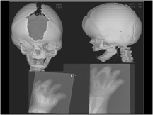
Figure 70.40 Apert's syndrome. (A,B) 3D CT surface-shaded display shows the wide open defect of the sagittal suture and brachycaphaly with bicoronal synostosis typical of Apert's syndrome. The coronal sutures appear fused and are ridged. (C,D) Plain radiographs of the hands show the ‘mitten hand’ appearance with syndactyly and shortened metacarpals
Slide 39
Figure 70.12 Child with skin lesions and right orbital cyst in oculocerebrocutaneous syndrome or Delleman's syndrome . (A) There is callosal agenesis with dorsal interhemispheric cysts. (B) The right cerebral hemisphere is dysplastic with thickening of the cortex and an indistinct grey–white matter junction (arrow). There is a cyst expanding the orbit with a small calcified globe seen inferiorly (arrowhead). (C) An associated Dandy–Walker posterior fossa malformation is also present
Slide 40
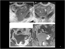
Figure 54.21 Endometrioma. Transvaginal US in sagittal (A) and coronal (B) planes demonstrates a complex cystic mass in the left ovary consistent with endometrioma (E in A, B). Although endometriomas can appear similar to haemorrhagic cysts, the irregular contour, homogeneity of the internal echoes and persistence over an extended period favours the diagnosis of endometrioma. Axial T1-weighted (C) and T1-weighted fat-suppressed MR (D) images in a different patient show multiple high signal intensity lesions within the left ovary (arrow, C), suggesting either endometriosis or haemorrhagic cysts. Note how fat suppression increases the conspicuity of haemorrhagic lesions and helps differentiate them from dermoids. Diagnosis of endometriosis was confirmed at surgery
Slide 41
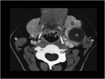
Figure 4-117 Second branchial cleft cyst. Computed tomography reveals a large thin-walled cystic mass (asterisk) anterior to the left sternocleidomastoid muscle (m) and posterior to the left submandibular gland (G)