Spotters 5

Slide 2
Adrenal mass & fatty sparing in liver.

Slide 3
MEDULLOBALSTOMA

Slide 4
CCA & BAND HETROTOPIA

Slide 5
Shows lungs hyperinflated.
The ascending aorta swings further to the right then expected, and the aortic knob is prominent.
The left border of the superior mediastinum has the appearance of an inverse 3.
Heart size is normal. 4th rib notchin on rt side

Slide 6
CM-2

Slide 7
CPA LIPOMA

Slide 8
Double patella syndrome:Double-layered patella is considered to be diagnostic for the recessive type of multiple epiphyseal dysplasia (MED).

Slide 9
AORTIC ROOT ECTASIA

Slide 10
Epidermoid. Enhanced scan shows a large, sharply marginated, low-attenuation, extra-axial sylvian mass (arrows).1

Slide 11
Stress fracture of the metatarsal (‘march fracture'). AP radiograph (A) demonstrates fluffy periosteal new bone along the distal shaft of the third metatarsal (arrow); the patient had foot pain for 16 days. In another patient, coronal STIR MRI (B) demonstrates increased signal in the distal second metatarsal diffusely, consistent with stress injury.

Slide 12
Craniopharyngioma. The rim-enhancing tumor contains dense calcification (straight arrows) and a large cystic component (curved arrows) that extends into the posterior fossa. Note the associated hydrocephalus.1

Slide 13
Discoid meniscus. (A) The plain film shows dishing of the
l ateral tibial plateau.

Slide 14
Hemangioblastoma in von Hippel-Lindau syndrome. (A) CT scan shows a cystic lesion (open arrows) with an enhancing nodule (closed arrow) in the left cerebellar hemisphere. (B) Vertebral arteriogram shows the vascular nodule (solid arrow) of the tumor with multiple feeding arteries (black arrowheads) and a large draining vein (open arrow).

Slide 15
Epidermoid. (A) Axial T1-weighted and (B) axial T2-weighted scans show an oblong mass (arrows) of slightly higher signal than CSF enveloping the basilar artery and extending around the pons.

Slide 16
Glomus jugulare tumor. Densely enhancing mass (arrow) that has eroded the osseous margins adjacent to the right jugular foramen.1

Slide 17
Hypertrophic osteoarthropathy secondary to pulmonary
neoplasm.
Hypertrophic
osteoarthropathy-exuberant
periosteal reaction of the radius
and ulna. In this patient,
changes in the bones of the
hands were minimal.
especially over distal phalanges and around joints. Fibrosis may
lead to contractures (Fig. 38.96). These changes may be preceded
or accompanied by a polyarthritis similar to rheumatoid arthritis in
at least 10% of patients. Osteoporosis, joint space narrowing, erosions
and subluxations may be seen. Changes of an erosive
arthropathy in association with calcification should suggest the
presence of progressive systemic sclerosis. This is a so-called `overlap syndrome' comprising a mixture of features
of rheumatoid arthritis, dermatomyositis, systemic lupus erythematosus
and progressive systemic sclerosis.
Osteoporosis, soft-tissue swelling and joint space narrowing are
found at affected joints. The distribution also may mimic rheumatoid
arthritis, but distal interphalangeal joints may be affected and the
peripheral arthropathy may be asymmetrical. Erosive change is not
This arthropathy does not involve synovium but causes capsular
fibrosis. Deformities of the hands and feet that are initially
reversible occur. Lateral deviation of the hands and feet may be
Hypertrophic osteoarthropathy. Generalised and symmetrical
diffuse increase in uptake is associated with thickening of the bony image at
isotope scanning.
seen when these parts are examined without weight-bearing. When
the hands are pressed down onto the cassette surface, the deformities
vanish. The joint spaces are mainly preserved. True erosions
are not

Slide 18
Sigmoid volvulus in a 64-year-old woman. (A) Massively dilated distended gas-filled loop of sigmoid colon (B) Contrast enema showing
the twisted sigmoid colon (bird of prey sign).
1.Lack of haustra in the margin 2 Left flank overlap sign 3 Liver overlap sign 4 Apex under left hemidiaphragm 5 Apex above 10th thoracic vertebra 6 Inferior convergence on left 7 Air:fluid ratio greater than 2:1 8Pelvic overlap sign

Slide 19
Rigler's sign of pneumoperitoneum. The bowel loops have a 'ghost-like' appearance due to gas both inside and outside making the wall more apparent.

Slide 20
Vein of Galen aneurysm. Contrast-enhanced scan shows dilatation of the vein of Galen and straight sinus (open arrows). Note the prominent feeding vessels of the choroid plexus (closed arrows) and the anterior cerebral arteries (thin arrows).

Slide 21
Rathke's cleft cyst. (A) Sagittal and (B) coronal T1-weighted images show an ovoid lesion of high intensity (arrow) in the middle to posterior portion of the pituitary fossa.

Slide 22
Cong abs of diaphragm

Slide 23
Ovarian ca mets

Slide 24
Choroid plexus cyst

Slide 25
Vagal paraganglioma (a) Contrast-enhanced axial CT image reveals intense enhancement of a left carotid space mass (m). (b) Lateral angiographic view obtained with a left CCA injection demonstrates the hypervascular mass (arrow) displacing both the ECA and ICA anteriorly.

Slide 26
Differential Diagnosis of unilateral skin thickening of the breast:
Lymphatic obstruction
Prior axillary lymph node dissection.
Primary breast cancer with spread to axillary nodes.
Other metastatic disease to axillary nodes.
Lymphoma.
Inflammatory breast carcinoma.
Mediastinal lymphatic blockage:
Sarcoid
Hodgkin Disease
Metastatic involvement
Inflammation
Acute mastitis/Abscess
Radiation Therapy

Slide 27
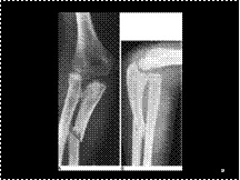
Galeazzi fracture-dislocation of the distal forearm. AP (A) and lateral (B) views of the distal arm demonstrate a displaced fracture of the radius and diastasis of the distal radioulnar joint, with ulnar dislocation

Slide 28
Carotid body tumor

Slide 29
Pulmonary agenesis / hypoplasia

Slide 30
CMF: There is a well defined lytic eccentric lesion in the metaphysis and proximal portion of the diaphysis of the right humerus. This has a well-defined zone of transition with normal bone and has the appearance of a line of demarcating sclerosis. The cortex on the medial side of the bone is thinned from below by the expanding lesion. The long axis of the lesion is parallel to the diaphysis. The skeleton is immature

Slide 31
Ccd
The skull vault is grossly normal for this age with the exception of the posterior fossa. The parietal and occipital bones are particularly widely separated. The bone margins of the foramen magnum are deficient. There is persistence of an accessory centre of ossification that is widely separated from the rest of the occipital bone and the condylar portion of basi-occiput. These normally join well-before birth. The scapulae are visible in this view, but the clavicles are absent.
The skull vault is grossly normal for this age with the exception of the posterior fossa. The parietal and occipital bones are particularly widely separated. The bone margins of the foramen magnum are deficient. There is persistence of an accessory centre of ossification that is widely separated from the rest of the occipital bone and the condylar portion of basi-occiput. These normally join well-before birth. The scapulae are visible in this view, but the clavicles are absent.

Slide 32
chondrosarcoma
There is a dense expansion of the bone on the medial side of the upper metaphysis of the right humerus with a base of thickened cortex. The lesion is also within the bone extending towards the diaphysis with a well-defined margin between higher and normal density. A soft tissue mass displaces the margin of the usual deltoid insertion. There is no visible lamellar periosteal reaction, although some irregular erosion of the lateral cortical margin is just visible on the lateral side below the greater tuberosity.
There is a dense expansion of the bone on the medial side of the upper metaphysis of the right humerus with a base of thickened cortex. The lesion is also within the bone extending towards the diaphysis with a well-defined margin between higher and normal density. A soft tissue mass displaces the margin of the usual deltoid insertion. There is no visible lamellar periosteal reaction, although some irregular erosion of the lateral cortical margin is just visible on the lateral side below the greater tuberosity.

Slide 33
MCU:
VUR, Dilated b/l ureters,rt PC system, ? VUR bcoz of dilated urethra

Slide 34
Bankhart injury of the inferior glenoid rim. AP (A) and axillary (B) radiographs in a patient who suffered an anterior shoulder dislocation show an irregularity in the inferior bony glenoid, consistent with a Bankhart fracture (arrow). The shoulder has been reduced. Axial CT (C) from the same patient shows the relationship of the fragment (arrow) to the glenoid.

Slide 35
Gastric volvulus can be divided into organoaxial, mesenteroaxial and mixed types. Organoaxial volvulus is the most common, where the stomach is rotated around its longitudinal axis (along a line extending from cardia to pylorus). In organoaxial volvulus the stomach can twist either anteriorly or posteriorly. The antrum moves from an inferior to superior position. In primary gastric volvulus the stabilizing ligaments of the stomach are too lax due to congenital or acquired causes. Secondary gastric volvulus occurs with a para-oesophageal hernia. Organoaxial volvulus is typically observed in adults with a large hiatus hernia and complications are generally rare.

Slide 36
(A), Radiograph: cecal volvulus. Radiograph from a patient with cecal volvulus shows a dilated cecum with no air distally in the colorectum. Convergence of the medial walls of the loop (black arrow) points to the right, a typical finding in cecal volvulus. (B), Barium enema: cecal volvulus. Barium examination demonstrates a bird's-beak deformity tapering at the point of volvulus (large white arrow). Note walls of dilated cecum (small white arrows).

Slide 37
Monteggia fracture-dislocation of the proximal forearm. AP (A) and lateral (B) views demonstrate an anteriorly angulated fracture of the proximal ulna and anterior dislocation of the radius relative to the capitellum.

Slide 38
Tarsal coalition occurs in approximately 1%. It typically presents in adolescence. 50% are bilateral (even if symptomatic on only one side). Frequently pes planus (flat foot) is a feature. Coalition may be: bony, fibrous (this case) or cartilaginous.
The commonest coalition is calcaneonavicular. This is best seen on an oblique projection.
In a bony coalition a 'bony bar' is observed. In a fibrous coalition irregularity and narrowing of the bony interfaces with sclerosis is identified (this case). If further imaging is required CT if the investigation of choice.

Slide 39
MECKEL'S DIVERT

Slide 40
AORTOCAVAL ANAST

Slide 41
Bipartite patella (A) is usually superolateral in location; the fragment is often smaller than the 'defect' in the patella. The edges are smooth and sclerotic (arrow).
In comparison, an acute fracture of the patella (B) demonstrates fragments that fit together like puzzle pieces; the edges are indistinct (arrow).

