Spotters 8
Slide 2
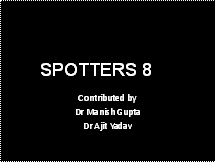
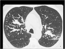
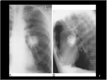
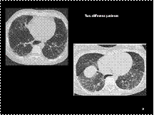
Chronic histoplasmosis. A 55 year old man. A PA chest radiograph demonstrates several well defined uniform-sized calcified nodules in both lungs with bilateral hilar and mediastinal lymph node calcification.
If unsuspected the disease may be diagnosed in retrospect years later by the appearance on chest radiograph of multiple calcified 3–4 mm in diameter sharply-marginated, round nodules and calcified lymph nodes in the hila and mediastinum. Occasionally a solitary, well defined nodule may form and is then termed a histoplasmoma.
Slide 3
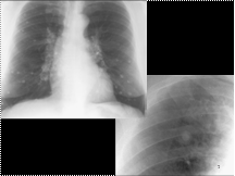
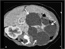
Benign ovarian teratoma with a mainly fatty component. There is a well-circumscribed, fatty mass in the lower pelvis with a peripheral soft tissue nodule. Even in the absence of calcific elements, the presence of fatty tissue is reliable enough to suggest a diagnosis of teratoma. The spectrum of CT findings in mature teratomas relates to their origin from three germ cell layers.
Slide 4
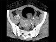
Galactocele:::more frequently occur after cessation of breast-feeding, when milk is retained and becomes stagnant within the breast
Slide 5
Allergic bronchopulmonary aspergillosis. HRCT of the upper lobes. Mucoid impactions are present within segmental and subsegmental dilated bronchi in the upper lobes. Small centrilobular linear branching opacities are seen in the periphery of the right upper lobe.
Slide 6
Lingular collapse. (A) Frontal view of isolated collapse of the lingular segments of the left upper lobe showing loss of clarity of the left heart border and a raised hemidiaphragm. (B) The similarity to a right middle lobe collapse can be appreciated on the lateral view.
Slide 7
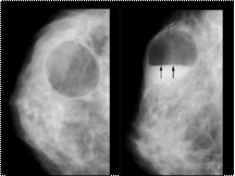
The lucent-centered calcifications are typical of fat necrosis
Slide 8
Bilateral lower lobe collapse. Bilateral triangular densities are seen with obscuration of the medial portions of the hemidiaphragms. The cause was mucous plugging.
Slide 9
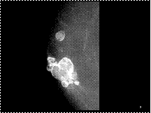
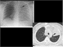
Hamartoma of the lung. (A,B) Round, completely smooth, hamartoma in a 57 year old asymptomatic man. There is typical coarse popcorn calcification in this lesion which is unusually large.
Slide 10
Luftsichel sign. (A) A left upper lobe collapse demonstrating paramediastinal lucency (arrow). (B) CT shows interposition of aerated lung between the collapse and the mediastinum (arrow). There is also a large right paratracheal node causing some distortion of the SVC.
Slide 11
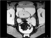
Ectopic ureterocele. The contrast-filled ureterocele (U) occupies the base of the bladder (BL). The low-attenuation rim represents the wall of the ureter. This patient had a duplicated left kidney with an obstructed upper pole moiety
Slide 12
Renal lymphoma
Slide 13
L renal lymphangioma
Slide 14
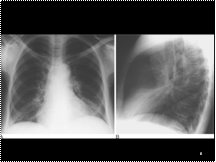
Left upper lobe collapse. Intravenous contrast-enhanced CT of left upper lobe collapse shows increased wedge-shaped density of the left upper lobe adjacent to the mediastinum. Note the displacement of the right lung across the midline anteriorly, resulting in retrosternal hyperlucency and increased clarity of the anterior ascending thoracic aorta on the lateral view.
Slide 15
Pancreatic calcifications in cystic fibrosis. A: Multiple parenchymal calculi are noted in the pancreatic tail (arrows) on this portal venous phase CT image. B: Numerous large calcifications along with fatty replacement of the pancreas (arrows) are noted in another patient.
Slide 16
An example of a fixed sliding hiatal hernia together with several B or Schatski rings.
Slide 17
Scleroderma. (A,B) Two patients with scleroderma showing ground-glass opacification in association with traction bronchiectasis and a fine reticular pattern. The pattern of fibrosis is closest to that of non-specific interstitial pneumonia. Note the dilated oesophagus in both examples.
Slide 18
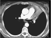
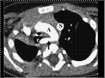
Left aortic arch with aberrant right subclavian artery. Axial CT image shows a left aortic arch (A) and an aberrant right subclavian artery (arrow) coursing posterior to the esophagus (e) and trachea. Note also a left superior vena cava (S).
Slide 19
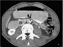
Superior mesenteric artery syndrome. The third portion of the duodenum is compressed as it passes between the aorta (A) and the superior mesenteric artery (arrow). Note the normal relationship of the superior mesenteric vein (arrowhead), to the right of the artery. The duodenum (D) proximal to the obstruction is dilated.
Slide 20
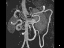
A contrast-enhanced MRA confirms mid aortic syndrome with focal occlusion of the juxtarenal aorta. The distal aorta is perfused by the mesenteric arteries. There is a grossly hypertrophied inferior mesenteric artery together with hypertrophied marginal artery and arc of Riolan.
Slide 21
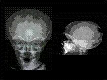
Parietal foramina
Slide 22
Annular Ca distal descending Colon
Slide 23
Polysplenia with situs inversus. Contrast-enhanced CT scan demonstrates multiple splenules (S) in the right upper quadrant posterior to the stomach (St). The liver (L) is left sided.

Slide 24
Pseudomembranous colitis. There is severe low-attenuation, mural thickening of the right and transverse colon. Intraluminal contrast agent is insinuating between thickened edematous folds in the transverse colon, producing the accordion sign (arrows). The left colon is fluid filled and shows mild wall thickening
Slide 25
EBSTEIN’S ANOMALY à CONGENITAL TRICUSPID REGURGITATION ? RA ENLARGEMENT, DECREASED FLOW IN PULMONARY ARTERY
Slide 26
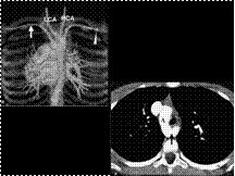
Right aortic arch with aberrant left subclavian artery. A: Axial CT scan demonstrates a right aortic arch (A) with the aberrant left subclavian artery (arrow) crossing the mediastinum posteriorly. B: Coronal 3D reconstruction (posterior view). The left subclavian artery (arrow) is the last vessel arising from the aorta. LCA, left common carotid artery; RCA, right carotid artery; right subclavian artery (arrowhead
Slide 27
Midgut malrotation and volvulus. A, B: Two axial CT scans of the upper abdomen in a patient with acute abdominal pain and vomiting shows clockwise swirling of the superior mesenteric vein (black arrow) and transverse duodenum (arrowhead) around the superior mesenteric artery. Also note the dilated proximal duodenum (D).
Slide 28
TAPVC / PAPVC
Slide 29
OM looser zone (pseudo#)
Slide 30
melorrhostosis
Slide 31
Rheumatoid arthritis. Characteristic erosion of the ulnar styloid process (arrow) by an adjacent tenosynovitis of the extensor carpi ulnaris tendon. Note the associated soft-tissue swelling.
Slide 32
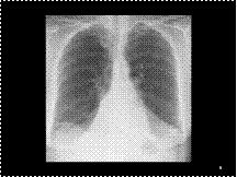
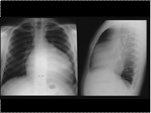
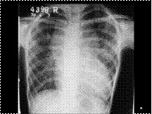
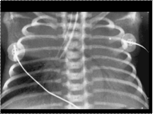
Malpositioned endotracheal tube. Inordinately low position of the endotracheal tube in the bronchus intermedius causes collapse of the right upper lobe and the entire left lung.
Slide 33
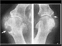
Gout. Two examples of typical rat-bite erosions about the first metatarsophalangeal joint (arrows). The cystlike lesions have thin sclerotic margins and characteristic overhanging edges.
Slide 34
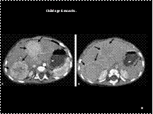
Hepatoblastoma. A: Arterial phase scanning shows two well-defined hyperattenuating tumors (arrows). The posterior one shows a mosaic enhancement pattern. B: On the late portal venous phase, the masses have become hypoattenuating. Note surrounding hyperdense capsules (arrows).
Slide 35
Fibrolamellar hepatocellular carcinoma. A: Late hepatic arterial phase scan shows a large heterogeneously enhancing mass in the left hepatic lobe. Note the central scar (arrow). B: In the portal venous phase, the tumor becomes isoattenuating with the liver. Again note the central hypoattenuating scar (arrow).
Slide 36
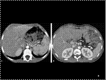
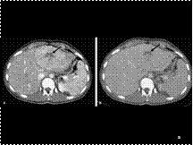
Hemochromatosis. The attenuation value of the liver is markedly higher than that of the spleen on this unenhanced CT in an adolescent girl who had multiple transfusions for sickle cell anemia.
Slide 37
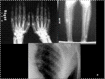
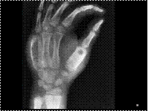
Hyperparathyroidism. (A) Dorsovolar radiograph of the hand of a 52-year-old man with hyperparathyroidism demonstrates the typical changes of this condition: increased bone radiolucency (osteopenia), subperiosteal resorption, loss of bony trabeculae, and tunneling of the cortices, which reflects a rapid bone turnover.
Slide 38
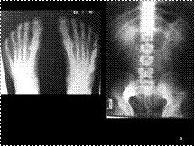
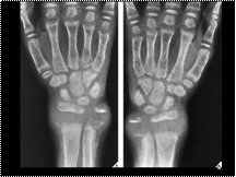
Rickets. (A), (B) Dorsovolar radiograph of both hands of an 8-year-old boy with untreated dietary rickets shows osteopenia of the bones, widening of the growth plates of the distal radius and ulna, and flaring of the metaphyses, all typical features of this condition
Slide 39
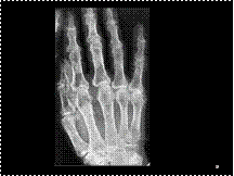
Tuberculosis of bone. Oblique film of the right hand of a 7-year-old boy with skeletal tuberculosis shows expansive fusiform lesions of the first and fifth metacarpals associated with soft-tissue swelling; there is no evidence of a periosteal reaction. Such diaphyseal enlargement secondary to tuberculosis is known as spina ventosa
Slide 40
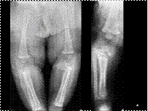
Congenital syphilis of bone. (A) Anteroposterior radiograph of the lower legs of a 7-week-old infant with congenital syphilis demonstrates characteristic periostitis affecting the femora and tibiae. In addition, destructive changes are evident in the medullary portion of the proximal tibiae. (B) Two months later, the infectious process has progressed, with destruction of the tibial metaphysis and marked periostitis. The characteristic erosion of the medial surface of the proximal tibial metaphysis is termed the Wimberger sign
Slide 41
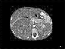
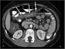
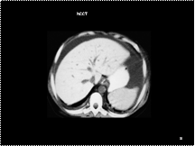
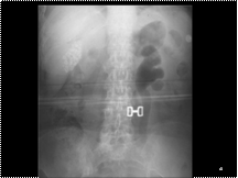
Emphysematous pyelonephritis. Patient with diabetes mellitus and sepsis. The left renal collecting system and ureter are distended and gas filled. There are also multiple dense gallstones in the gallbladder.